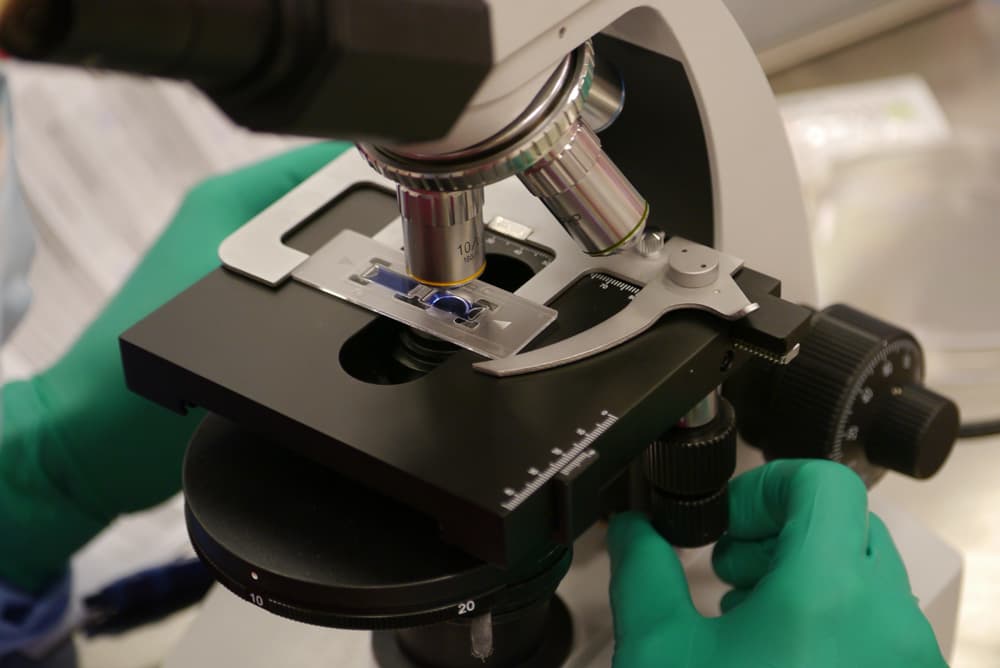
Regenerative micrograft preparation at London Cartilage Clinic

The evidence

Mytocel MSK evidence: the studies behind the technology

A plain-English look at the published evidence behind the autologous micrografting technology used in Mytocel MSK, and an honest note on what varies by joint.

Quick answer

The evidence behind Mytocel MSK is evidence for the technology it is built on. Autologous micrografting has been studied in joint osteoarthritis by Marcarelli and colleagues, Tsoukas and colleagues, Helito and colleagues, with further work by Codorean and by Lacza. These studies test the technology, and outcomes vary by joint. The wider track record belongs to the technology, not to London Cartilage Clinic’s own numbers.

What we mean by evidence

Mytocel MSK is the musculoskeletal application of an established autologous micrografting technology, made by Remedi and built on the Regenera Activa system. Taking a small sample of a person’s own tissue and preparing it into living micrografts is a well-studied way to support natural repair, and it has been used across other fields of medicine for years.

The sensible way to read the evidence is to be clear about what it tests. The published studies below examine the micrografting technology, often in the knee. That is encouraging, but it is not a promise about every joint. Professor Lee is careful to separate what the technology has shown from what is realistic for your particular joint, which is judged on examination and imaging.

The studies behind the technology

A selection of the published work on autologous micrografting, described plainly so you can see what each one is and is not.

Marcarelli and colleagues

A three-year follow-up looking at autologous micrografting in knee osteoarthritis, one of the longer windows reported for the technology.

Tsoukas and colleagues

A prospective observational study of the micrografting technique, following patients forward rather than looking back at records.

Helito and colleagues

A clinical pilot applying the micrografting technology, an early-stage look at how it performs in practice.

Codorean and Lacza

Further work adding to the body of evidence on autologous micrografting, from separate research groups.

These studies examine the micrografting technology rather than every joint it can be used for. Results vary by joint, and Professor Lee will set realistic expectations for yours.

The technology’s track record

These figures describe the wider micrografting technology across medical fields. They are not London Cartilage Clinic’s own case numbers.

450,000+

procedures worldwide with the micrografting technology

80+

scientific publications behind the technology

10+ years

of clinical use across medical fields

Being honest about what varies

Mytocel MSK works with your own biology, so a general figure cannot promise a set result in your joint. Outcomes vary with the pattern and extent of wear, the joint being treated, and the individual. The evidence supports a genuine effect from your own repair cells, and it is the reason Mytocel MSK is offered on its own for earlier wear and as the cell component inside NanoACi, Professor Lee’s fuller injection protocol.

Mytocel MSK is from £3,000, the same across joints, confirmed at your consultation. The most useful next step is an imaging review, so the evidence can be applied to your joint rather than to joints in general.

Autologous micrografts prepared and checked at London Cartilage Clinic
The autologous micrografting technology behind Mytocel MSK, studied across other fields of medicine.

The published evidence

Mytocel MSK is built on autologous cartilage micrografting technology that has its own body of research. Here are the key studies, each with a downloadable summary and, where published, a link to the paper itself.

Cover of the study: Cartilage Micrografts as a Novel Non-Invasive and Non-Arthroscopic Autograft Procedure for Knee Chondropathy: Three-Year Follow-Up Study
Peer-reviewed

Cartilage Micrografts as a Novel Non-Invasive and Non-Arthroscopic Autograft Procedure for Knee Chondropathy: Three-Year Follow-Up Study

A single application of autologous cartilage micrografts improved patients’ quality of life and increased cartilage thickness on MRI three years later.

Marcarelli M, Zappia M, Rissolio L, Baroni C, Astarita C, Trovato L, Graziano A. Journal of Clinical Medicine. 2021;10(2):322.

Cover of the study: Prospective Observational Study of a Non-Arthroscopic Autologous Cartilage Micrografting Technology for Knee Osteoarthritis
Peer-reviewed

Prospective Observational Study of a Non-Arthroscopic Autologous Cartilage Micrografting Technology for Knee Osteoarthritis

One intra-articular injection of auricular cartilage micrografts improved pain and function in early-to-moderate knee osteoarthritis.

Tsoukas D, Muntean I, Simos C, Sabido-Vera R. Bioengineering. 2023;10(11):1294.

Cover of the study: Efficacy of Autologous Micrografting Technology in Managing Osteoarthritis Pain: A Pilot Study
Peer-reviewed

Efficacy of Autologous Micrografting Technology in Managing Osteoarthritis Pain: A Pilot Study

A clinical pilot found autologous micrografts reduced osteoarthritis pain, supporting the technology as a minimally invasive option for joint pain.

Helito CP, Pessei V, Zaniboni C, Muntean I. Bioengineering. 2024;11(11):1119.

Cover of the study: Single-Stage Knee Cartilage Repair Using Auricular Chondroprogenitor Micrografts with a Tri-Layer Scaffold: 12-Month Outcomes
Preliminary data

Single-Stage Knee Cartilage Repair Using Auricular Chondroprogenitor Micrografts with a Tri-Layer Scaffold: 12-Month Outcomes

At twelve months, combining auricular chondroprogenitor micrografts with a scaffold improved KOOS scores, presented as a single-stage alternative to two-stage cartilage surgery.

Codorean I, et al. Preliminary clinical data (not yet a peer-reviewed publication).

Cover of the study: Autologous Cartilage Micrograft Transplantation and the Joint Inflammatory Environment
Preliminary data

Autologous Cartilage Micrograft Transplantation and the Joint Inflammatory Environment

Auricular cartilage micrograft injection was associated with a reduction in the inflammatory signals (IL-1β, IL-6, IL-8, TNFα) that drive cartilage breakdown and joint pain.

Lacza Z, et al. Preliminary data (not yet a peer-reviewed publication).

Downloads are study summaries of the autologous micrografting technology, provided by the manufacturer. Peer-reviewed studies link to their published home; the figures describe the technology across published research, not London Cartilage Clinic’s own case numbers.

consulting-in-office-with-pen

Frequently Asked Questions

Is there evidence that Mytocel MSK works?

There is a published body of work behind the autologous micrografting technology Mytocel MSK is built on, including a three-year follow-up in knee osteoarthritis by Marcarelli and colleagues, a prospective observational study by Tsoukas and colleagues, a clinical pilot by Helito and colleagues, and further work by Codorean and by Lacza. Professor Lee will explain what this evidence does and does not yet show for your particular joint.

What do the studies actually measure?

The studies test the underlying micrografting technology, made by Remedi and built on the Regenera Activa system, rather than every individual joint it can be used for. That is an honest distinction: strong evidence for the technology is not the same as a guaranteed result in your specific joint, which is why your imaging review sets realistic expectations.

Do the 450,000 procedures refer to London Cartilage Clinic?

No. Figures such as 450,000+ procedures worldwide, 80+ publications and more than ten years of clinical use describe the wider micrografting technology across medical fields. They are not London Cartilage Clinic’s own case numbers, and we are careful not to present them as such.

Will Mytocel MSK give me the same result as the studies?

Outcomes vary by joint, by the pattern and extent of wear, and by the individual. The evidence points to a genuine biological effect from your own repair cells, but no study can promise a set result for your joint. What is realistic for you is judged on examination and imaging at your consultation.

How does the evidence for Mytocel MSK compare with NanoACi?

Mytocel MSK is the cell component inside NanoACi, Professor Lee’s injection protocol, so the two share the same micrografting biology. NanoACi adds a ChondroFiller scaffold and PRF alongside those cells. See the NanoACi evidence page for how that fuller protocol is framed.

Is Mytocel MSK safe?

Because it uses a small sample of your own tissue, prepared chair-side and injected in one session, it avoids the rejection and donor-tissue concerns of foreign material, and nothing is grown in a laboratory over weeks. The usual considerations of any joint injection apply, which Professor Lee will go through with you at consultation.

Still have more specific concerns?

Free Discovery Call

Options that our doctors may discuss include NanoACi, ChondroFiller, Mytocel MSK, joint replacement, established conservative care or surgery, depending on examination and imaging.

See whether the evidence applies to your joint

The only way to know what Mytocel MSK could achieve for you is an imaging review. Talk it through first on a free discovery call, or book a consultation with Professor Lee and have your imaging reviewed.

London Cartilage Clinic

Latest Insights

Clinical updates, cartilage treatment guidance, and recovery-focused articles from our specialist team.

Privacy & Cookies Policy