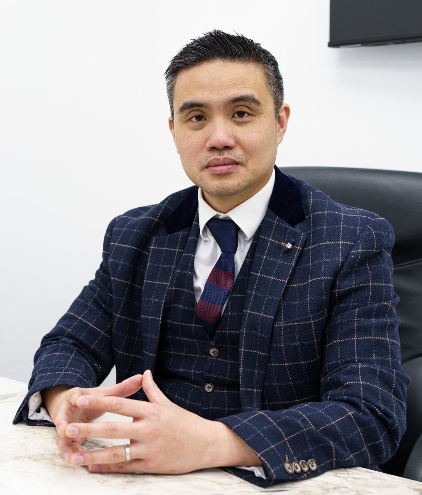
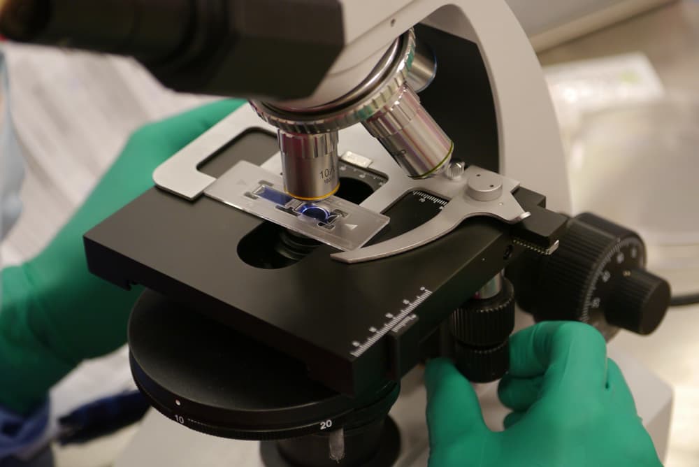
Professor Lee reviewing cartilage research and imaging

The evidence

The evidence behind NanoACi

NanoACi brings together three of your own tissues, each with its own published research. Here is what the evidence for each component shows, and an honest view of what the combined protocol does and does not yet prove.

Quick answer

The strongest evidence sits with each of NanoACi’s three components. The auricular micrografting technology behind the Mytocel MSK step, the ChondroFiller scaffold and PRF each have their own research. NanoACi is Professor Lee’s protocol for delivering them together in one session, so outcomes for the combination are reported case by case. We keep each component’s evidence labelled as that component’s, not as NanoACi’s own.

Three components, three bodies of evidence

NanoACi is an injection protocol Professor Paul Lee performs. In a single session he delivers three things that come from your own body: your cartilage cells (prepared with the Mytocel MSK micrografting technique), a ChondroFiller scaffold, and PRF. It is a technique, not a product or a device, and it does not create a new injectable. That matters for reading the evidence, because the research belongs to the components, and it is fairest to weigh it that way.

What each component shows

The research, component by component

The cells

The Mytocel MSK step

The cartilage cells are prepared with the Mytocel MSK auricular micrografting technique. The technology behind that step has been studied in joint osteoarthritis by Marcarelli and colleagues (a three-year follow-up), Tsoukas and colleagues (a prospective observational study) and Helito and colleagues (a clinical pilot), with further work by Codorean and by Lacza. This is evidence for the micrografting technology, not for NanoACi as a whole.

The scaffold

ChondroFiller

ChondroFiller has its own body of evidence for supporting cartilage repair, and it is already used at the clinic for that purpose. It is the soil the cells settle on and rebuild from.

The growth signal

PRF

Platelet-rich fibrin, prepared from your own blood on the day, is well established in regenerative medicine. It adds the growth signals that help new tissue take.

The honest position

As with any newer combination, the strongest published evidence sits with each component in its own right. Bringing the three together in one session is Professor Lee’s protocol, and outcomes for that pairing are reported case by case rather than from a large trial of NanoACi as a single named treatment. We think it is more useful to say that plainly than to borrow one component’s research to speak for the whole.

So the studies of the auricular micrografting technology stay labelled as the Mytocel MSK step’s evidence, the ChondroFiller research stays the scaffold’s, and PRF’s standing stays its own. Cartilage cell therapy improves symptoms and joint function; long-term regrowth of new cartilage continues to be studied. Professor Lee will walk you through what the evidence does and does not yet show for your particular joint.

The micrografting technology behind the Mytocel MSK step has been used in over 450,000 procedures worldwide across medical fields and is supported by more than 80 scientific publications over more than ten years. Those figures describe the wider technology’s track record, not London Cartilage Clinic’s own case numbers.

Cartilage cell therapy under study at London Cartilage Clinic
Each component of NanoACi carries its own evidence; the combined protocol is reported case by case.

The evidence behind the cells

NanoACi's cell component is prepared with the Mytocel MSK micrografting technique, which draws on the auricular cartilage micrografting research below. ChondroFiller and PRF each have their own separate evidence.

Cover of the study: Cartilage Micrografts as a Novel Non-Invasive and Non-Arthroscopic Autograft Procedure for Knee Chondropathy: Three-Year Follow-Up Study
Peer-reviewed

Cartilage Micrografts as a Novel Non-Invasive and Non-Arthroscopic Autograft Procedure for Knee Chondropathy: Three-Year Follow-Up Study

A single application of autologous cartilage micrografts improved patients’ quality of life and increased cartilage thickness on MRI three years later.

Marcarelli M, Zappia M, Rissolio L, Baroni C, Astarita C, Trovato L, Graziano A. Journal of Clinical Medicine. 2021;10(2):322.

Cover of the study: Prospective Observational Study of a Non-Arthroscopic Autologous Cartilage Micrografting Technology for Knee Osteoarthritis
Peer-reviewed

Prospective Observational Study of a Non-Arthroscopic Autologous Cartilage Micrografting Technology for Knee Osteoarthritis

One intra-articular injection of auricular cartilage micrografts improved pain and function in early-to-moderate knee osteoarthritis.

Tsoukas D, Muntean I, Simos C, Sabido-Vera R. Bioengineering. 2023;10(11):1294.

Cover of the study: Efficacy of Autologous Micrografting Technology in Managing Osteoarthritis Pain: A Pilot Study
Peer-reviewed

Efficacy of Autologous Micrografting Technology in Managing Osteoarthritis Pain: A Pilot Study

A clinical pilot found autologous micrografts reduced osteoarthritis pain, supporting the technology as a minimally invasive option for joint pain.

Helito CP, Pessei V, Zaniboni C, Muntean I. Bioengineering. 2024;11(11):1119.

Cover of the study: Single-Stage Knee Cartilage Repair Using Auricular Chondroprogenitor Micrografts with a Tri-Layer Scaffold: 12-Month Outcomes
Preliminary data

Single-Stage Knee Cartilage Repair Using Auricular Chondroprogenitor Micrografts with a Tri-Layer Scaffold: 12-Month Outcomes

At twelve months, combining auricular chondroprogenitor micrografts with a scaffold improved KOOS scores, presented as a single-stage alternative to two-stage cartilage surgery.

Codorean I, et al. Preliminary clinical data (not yet a peer-reviewed publication).

Cover of the study: Autologous Cartilage Micrograft Transplantation and the Joint Inflammatory Environment
Preliminary data

Autologous Cartilage Micrograft Transplantation and the Joint Inflammatory Environment

Auricular cartilage micrograft injection was associated with a reduction in the inflammatory signals (IL-1β, IL-6, IL-8, TNFα) that drive cartilage breakdown and joint pain.

Lacza Z, et al. Preliminary data (not yet a peer-reviewed publication).

Downloads are study summaries of the autologous micrografting technology, provided by the manufacturer. Peer-reviewed studies link to their published home; the figures describe the technology across published research, not London Cartilage Clinic’s own case numbers.

consulting-in-office-with-pen

Frequently Asked Questions

Is there evidence that NanoACi itself works?

The honest answer is that the strongest evidence sits with each of the three components, not yet with the combined protocol as a single named treatment. The auricular micrografting technology behind the Mytocel MSK step, the ChondroFiller scaffold and PRF each have their own published research. Outcomes for the components delivered together are reported case by case, and Professor Lee will explain what does and does not yet apply to your joint at consultation.

What research supports the cell step?

The cells are prepared with the Mytocel MSK auricular micrografting technique. The technology behind it has been studied in joint osteoarthritis by Marcarelli and colleagues (a three-year follow-up), Tsoukas and colleagues (a prospective observational study) and Helito and colleagues (a clinical pilot), with further work by Codorean and by Lacza. That is evidence for the micrografting technology, not for NanoACi as a whole. See the Mytocel MSK evidence page.

Is there evidence for the ChondroFiller scaffold?

Yes. ChondroFiller has its own body of evidence for supporting cartilage repair and is already used at the clinic in its own right. In NanoACi it provides the structure your own cells settle on and rebuild from.

What about the PRF?

Platelet-rich fibrin is well established in regenerative medicine. It is prepared from a small sample of your own blood on the day of treatment and adds the growth signals that help new tissue take.

What do the figures of 450,000 procedures and 80 publications refer to?

Those numbers describe the wider auricular micrografting technology behind the Mytocel MSK step: more than 450,000 procedures worldwide across medical fields, over 80 scientific publications and more than ten years in use. They are the technology’s track record. They are not London Cartilage Clinic’s own case numbers, and they should not be read as outcomes for NanoACi as a whole.

Will Professor Lee tell me what the evidence shows for my joint?

Yes. At consultation Professor Lee sets out what the component evidence supports, what is still being studied, and how that applies to your imaging and your particular joint. Start with a free discovery call, then book a consultation. See the NanoACi treatment page or check whether NanoACi is right for you.

Still have more specific concerns?

Free Discovery Call

Options that our doctors may discuss include NanoACi, ChondroFiller, Mytocel MSK, joint replacement, established conservative care or surgery, depending on examination and imaging.

Talk through the evidence for your joint

A free discovery call is the easiest first step. Professor Lee will explain what the component evidence supports and what is still being studied, then review your imaging so any plan is grounded in your own scan.

London Cartilage Clinic

Latest Insights

Clinical updates, cartilage treatment guidance, and recovery-focused articles from our specialist team.

Privacy & Cookies Policy